CRL Reports: Conserving the human skeleton found aboard La Belle
Conserving the human skeleton found aboard La Belle
La Salle Shipwreck Project
Texas Historical Commission
Throughout each year, the Conservation Research Laboratory conserves material from a number of different archaeological projects. The purpose of these CRL reports is to showcase the conservation procedures used to treat some of the more interesting archaeological material. The conservation of the human skeleton is presented in this report. The Belle, one of the ships of French explorer Robert Cavelier, Sieur (Lord) de La Salle, was lost in Matagorda Bay, Texas in 1686. It was excavated by the Texas Historical Commission.
Skeletal material always creates a lot of attention and interest among the public, and the articulated remains of an individual found on a forward deck in the hold of the Belle was no exception. Extensive news coverage was given to this particular find.
The human skeleton from the Belle was discovered by archaeologists from the Texas Historical Commission (THC) on October 31, 1996. It was delivered to the Conservation Research Laboratory soon after it was removed. In the last few years, it has undergone conservation, various analyses, and we even managed to reconstruct the face.
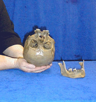
Due to the anaerobic burial conditions, the skeleton was in excellent condition. In fact, the burial environment was so good that a large amount of tendon tissue remained on various bones and, most significantly, a large portion of the brain was preserved in the cranium. Since the cranium contained brain tissue, it had to be kept inverted to keep it from falling out of the foramen magnum. This is the reason that the conservator in the image to the right is holding the skull in an inverted position.
Drs. Donny Hamilton and Wayne Smith decided that the Belle skeleton would be an excellent case study to demonstrate to the lay public the technology now available that can assist archaeologists in interpreting archaeological data. The project described here is the result of a number of specialists and technologies, all coming together to present an in-depth view of conservation and archaeological analysis. The project was undertaken with the permission of the Texas Historical Commission.
BONE CONSERVATION
In most instances, the conservation of bone is a fairly straightforward process. In the case of the Belle skeleton, the first problem to be confronted resulted from the fact that bone is porous – having being submerged in sea water for over three centuries, the skeleton had absorbed soluble salts. These salts had to be removed to stabilize the bones. To do this simply meant that the bones had to be rinsed in running tap water, followed by successive baths of de-ionized water. The bones were then dried by putting them through successive baths of ethanol and acetone. The skeleton was then consolidated in polyvinyl acetate V15 in acetone under a vacuum.
As the bones were undergoing the water rinses, tissue samples were removed and placed in small vials filled with 50 percent water / 50 percent ethanol. Each vial was labeled as to location and possible type of tissue sample. Selective samples of the brain material were collected in the same manner. Some of the samples were taken to the Archaeological Preservation Laboratory for conservation treatment with silicone oils. This is to determine whether silicone treatment can be used to preserve tissue for indefinite periods without adversely affecting the DNA in the tissue samples. A final decision has not been made on how best to treat the remaining brain tissue.
The skull with the enclosed brain, as well as the mandible, are currently being stored in 50 percent water / 50 percent ethanol bath until it can be determined what tests might be run on them.
FACIAL RECONSTRUCTION TECHNOLOGY
In connection with a Paleo-Indian facial reconstruction project in 1996, the Conservation Research Laboratory staff members were contacted by representatives from CyberForm International (CFI) in Richardson, Texas, informing us of a new casting procedure called stereolithography that could produce an exact replica of the skull — both exterior and interior features — without making a mold. At the time, we did not have a project in mind for the proposed technology; however, when the individual was found on the Belle, Dr. Wayne Smith got in touch with CFI and arranged for the project. The facial reconstruction project described here required three major steps:
- a computer tomograph scan
- a stereolitography cast of the skull
- a skilled technician to model the face in clay and make the molds
COMPUTER TOMOGRAPHY SCAN
The first step in making a replica of the skull from the Belle was to have it scanned using a computer tomography (CT) scan. In March 1997, the Texas Scottish Rite Hospital for Children in Dallas, Texas, graciously volunteered their technicians and their newly installed Phillips Tomograph AV to do the scan. The CT imaging technicians at the hospital made very fine, detailed 3-D digital images of the skull. Significantly, the CT scan showed the extent of the remaining brain tissue and provided us with the first volumetric indication of the amount of brain present. Despite various media accounts, no evidence of a pointed metal object was found in the skull. A small pebble was found in the cranium that probably accounts for the shadow that showed up on the CT scan.
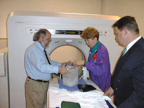
Above left, Wayne Smith of Texas A&M University, Lois Liehman, the computer tomograph technician from the Texas Scottish Rite Hospital for Children, and Marc McAllister of CyberForm International prepare the skull for the CT with a new Phillips Computer Tomograph AV. Above right, the scanned skull.
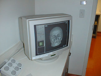
The CT scan produced a digital 3-D image that recorded all of the diagnostic features of the skull. These data, which are stored on a computer disk, are a permanent part of the archaeological record of the skeleton. The digital image can be viewed at any time and from any perspective (see below). It can be also used to make additional casts of the skull in the future if the need arises.
 |
 The gray shadow is formed by the liquid-filled plastic bag containing the skull. The cloudy area inside the cranium is produced by the preserved brain tissue. The white area represents a cross section through the bone of the skull. |
 Note the bad dentition, missing teeth, and abscesses in the mandible bone. |
STEREOLITHOGRAPHY CAST
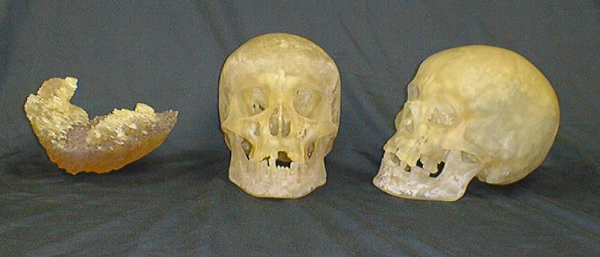
After securing the CT scan, Marc McAllister of CyberForm International took the digital data, and by a casting process called stereolithography, made two exact resin replicas of the skull (below). To the left of the two skulls is a resin cast of the brain in the cranium, which corresponds to the cloudy area in the center CT digital image above. The resin brain cast (below, left) clearly shows the volume of brain remaining in the skull.
See the Facial Reconstruction